
The knee stands as one of the human body’s largest and most robust joints, playing a pivotal role in facilitating multi-directional movement. Acting as the connecting link between the thigh bone (femur) and the leg bone (tibia), the knee relies heavily on ligaments for its stability and functionality.
Ligament Knee Injuries Overview
Ligaments, composed of dense and resilient connective tissue, are essential for upholding the stability of the knee joint. They enable various movements such as walking, bending, running, and pivoting. The knee is stabilized primarily by four key ligaments:
Anterior Cruciate Ligament (ACL) and Posterior Cruciate Ligament (PCL): Situated inside the knee, these ligaments intersect to form an “X” shape, preventing excessive forward and backward movement of the knee joint.
Medial Collateral Ligament (MCL) and Lateral (Fibular) Collateral Ligament (FCL or LCL): Located on the sides of the knee, these ligaments serve to limit side-to-side motion of the joint.
Ligament tears are a common occurrence, particularly among athletes engaged in activities such as soccer, football, basketball, skiing, and gymnastics. While single ligament injuries like ACL tears or MCL tears are prevalent, severe trauma or force applied to the knee can lead to multiple ligament injuries. Events like motor vehicle accidents or high-impact collisions during sports can result in complex knee injuries.
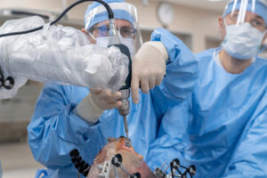
In cases where multiple knee ligaments are injured, additional complications such as knee dislocation or fractures may arise. Such complex injuries often necessitate surgical intervention by an orthopedic surgeon to reconstruct the damaged ligaments.
For more information or assistance regarding knee ligament injuries and their treatment options, individuals are encouraged to consult with Dr. Ravi Teja Rudraraju at https://drraviteja.com.
Treatment for Multi-Ligament Knee Injuries
A thorough clinical examination combined with stress x-rays plays a crucial role in diagnosing multi-ligament knee injuries, particularly in cases of chronic injury or traumatic events where determining the source of side-to-side laxity is challenging. In such scenarios, evaluating the patient’s alignment, especially in chronic cases, and obtaining a high-quality MRI scan are essential to identify any accompanying cartilage or meniscus injuries.
Initially, non-operative treatment may be recommended, especially in cases of concurrent ACL and medial-sided knee injuries, allowing the MCL to heal first. Similarly, for grade I or grade II injuries of the medial or lateral knee structures alongside cruciate ligament injuries, conservative measures may suffice. However, when significant instability persists, Dr. Ravi Teja Rudraraju typically advises a concurrent multiple ligament reconstruction. The primary goal of such reconstructive procedures is to achieve secure and anatomically positioned ligament reconstruction, enabling early range of motion to prevent stiffness and knee scarring, ultimately enhancing patient function and minimizing the risk of osteoarthritis.
Post-Operative Care
Following multi-ligament knee injury reconstruction, a well-guided physical therapy program is imperative for optimal recovery. Patients must aim to regain full knee extension promptly, focusing on quadriceps activation and edema control. Achieving a range of motion from 0 to 90 degrees within the first two weeks is crucial, followed by further progression between weeks two and six. Typically, patients are advised to refrain from weight-bearing for the initial six weeks postoperatively.
Recovery from complex knee injuries necessitates a comprehensive rehabilitation period of 9-12 months before returning to full activities. Allowing adequate time for ligament healing and preventing premature return to activities can reduce the risk of graft failure or fatigue, as well as prevent reinjury to the operated or contralateral knee. For more information on multi-ligament knee injuries and their treatment, individuals are encouraged to visit https://drraviteja.com.
Post-Operative Care:
Following multi-ligament knee injury reconstruction, a well-guided physical therapy program is imperative for optimal recovery. Patients must aim to regain full knee extension promptly, focusing on quadriceps activation and edema control. Achieving a range of motion from 0 to 90 degrees within the first two weeks is crucial, followed by further progression between weeks two and six. Typically, patients are advised to refrain from weight-bearing for the initial six weeks postoperatively.
Recovery from complex knee injuries necessitates a comprehensive rehabilitation period of 9-12 months before returning to full activities. Allowing adequate time for ligament healing and preventing premature return to activities can reduce the risk of graft failure or fatigue, as well as prevent reinjury to the operated or contralateral knee. For more information on multi-ligament knee injuries and their treatment, individuals are encouraged to visit https://drraviteja.com.
Have you sustained a multi-ligament knee injury?
There are two ways to initiate a consultation with Dr. Ravi Teja Rudraraju
You can provide current X-rays and/or MRIs for a clinical case review with with Dr. Ravi Teja Rudraraju
You can schedule an office consultation with Dr. Ravi Teja Rudraraju
Frequently Asked Questions
A multiple ligament knee injury occurs when at least two of the four main ligaments of the knee are injured. These ligaments include the ACL, PCL, MCL (along with its components), and the posterolateral corner of the knee (including the FCL/LCL, popliteus tendon, and popliteofibular ligament). Severe cases may involve knee dislocation.
Diagnosing multiple ligament knee injuries involves a thorough history review with the patient to identify any dislocation events or sensations of knee instability. A complete physical examination is crucial, including specific tests to assess the integrity of various ligaments such as the Lachman test for the ACL, the posterior drawer test for the PCL, and valgus/varus stress tests for the MCL and lateral structures. Additionally, imaging modalities like MRI scans and stress x-rays may be necessary for a comprehensive evaluation.
Optimal treatment for multiple ligament knee injuries involves early surgical intervention with anatomic-based reconstructions of the injured ligaments. This approach aims to restore knee stability and function, minimize the risk of stiffness and scarring, and prevent long-term complications such as osteoarthritis. Physical therapy focusing on early range of motion is crucial for successful rehabilitation.
The current standard of care for multi-ligament knee injuries emphasizes early surgical reconstruction of the affected ligaments using anatomical techniques. This approach, coupled with early postoperative physical therapy and rehabilitation, has demonstrated superior outcomes compared to traditional methods. Dr. Ravi Teja Rudraraju’s team at https://drraviteja.com follows these modern principles to ensure optimal patient care and outcomes.
Knee pseudo-instability refers to the perception of knee instability caused by factors such as arthritis affecting one compartment of the knee. Patients may experience a wobbling sensation during movement, resembling true knee instability. Careful clinical examination and imaging studies are essential to differentiate between true ligamentous laxity and pseudo-instability, guiding appropriate management strategies. For more information on knee instability and its treatment, individuals can visit https://drraviteja.com.
Yes, knee instability can occur without significant pain. Pain in the knee is often associated with conditions like arthritis or meniscus tears, while instability can be caused by ligament injuries such as PCL tears. Despite the absence of pain, untreated instability may lead to gradual cartilage wear and arthritis over time. Thus, individuals experiencing knee instability without pain should consider seeking evaluation and treatment to prevent future complications such as arthritis.
Side-to-side knee instability occurs when there is damage to structures on either side of the knee joint, leading to a sensation of the joint moving laterally during movement. This instability can be felt during activities such as planting the foot or walking, and it may indicate injuries to ligaments such as the MCL or lateral structures. However, in some cases, perceived instability may be due to arthritic changes rather than true ligamentous laxity.
Knee instability while descending stairs can be caused by various factors. Weakness in the quadriceps muscles may lead to a sensation of the knee giving way due to inadequate support. True instability, where the knee feels like it’s sliding forward during descent, is often associated with PCL tears. Patients with chronic PCL tears may benefit from wearing a dynamic brace to stabilize the knee and confirm the diagnosis before considering surgical reconstruction.
Yes, knee instability can indirectly contribute to back pain. Limping due to knee instability can alter the mechanics of the sacroiliac joint and lead to weakness in the hip abductors. This weakness, along with changes in gait, can result in discomfort in the lower back region. Addressing the underlying cause of knee instability and implementing hip abduction strengthening exercises can help alleviate associated back pain.
Various imaging modalities are used to assess multi-ligament knee instability. X-rays, including long-leg views, help evaluate alignment and detect fractures. Stress x-rays assess ligamentous laxity, while MRI scans provide detailed information about ligament tears, cartilage status, and associated injuries. These imaging studies aid in accurate diagnosis and treatment planning for patients with multi-ligament knee injuries. For comprehensive evaluation and management of such injuries, individuals can consult with Dr. Ravi Teja Rudraraju and visit https://drraviteja.com.