

What is the PCL (Posterior Cruciate Ligament)?
If you are experiencing discomfort behind your knee, it could potentially indicate a PCL tear or strain.
The posterior cruciate ligament (PCL) is situated at the back and middle of the knee. It is one of the key ligaments that connect the femur (thighbone) to the tibia (shinbone). Renowned for its strength, the PCL is less prone to injury, accounting for approximately 3-37% of all knee injuries.
PCL Tear Symptoms
Tears in the posterior cruciate ligament may lead to the following symptoms:
In many cases, a posterior cruciate ligament injury occurs due to a forceful impact, often seen in sports-related trauma. Causes of PCL injuries may include a bent knee striking a hard surface (such as a car dashboard in an accident or a hockey player colliding with a goalpost) or a football player falling onto a bent knee.
PCL Diagnosis
Dr. Ravi Teja Rudraraju will conduct a thorough clinical examination to identify signs of a torn PCL. This assessment may be complemented by x-rays, posterior knee stress x-rays while kneeling, and frequently, an MRI scan to assess the extent of the injury and any accompanying injuries. Diagnosing a PCL tear early is crucial to promote healing in a stable position rather than in an elongated and nonfunctional state. While MRI results are valuable for evaluating acute injuries, they may not effectively diagnose chronic PCL injuries, where the ligament may have healed in an elongated position, necessitating stress x-rays for accurate diagnosis of the tear extent.
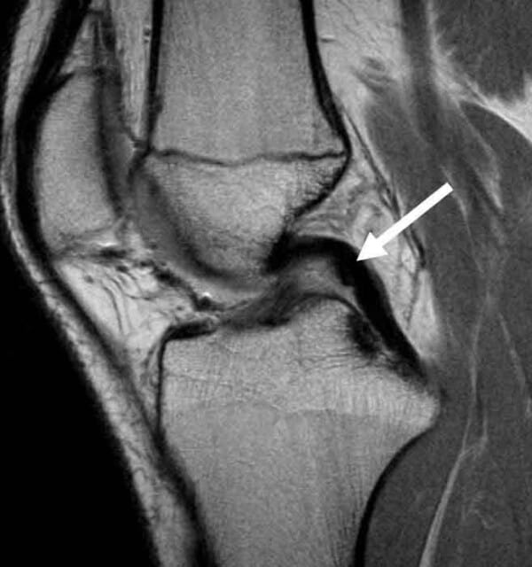
Sagittal MRI scan displaying the typical pathway of the PCL from the tibia to the femur. It’s noteworthy that the attachment point on the tibia is situated at the lower portion of the PCL facet.
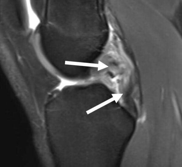
Sagittal MRI scan illustrating a PCL tear. It’s evident that there is a disruption in the continuity of the PCL fibers, which typically appear dark in signal and thick.
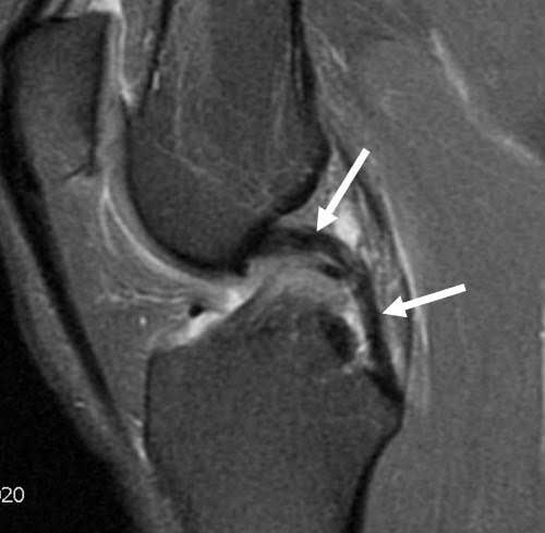
MRI sagittal scan depicting a chronic PCL injury that has healed in a loose and nonfunctional position, despite being inaccurately interpreted as normal in the MRI report.
PCL Injury Test
The diagnosis of a torn posterior cruciate ligament relies on evaluating the patient’s posterior knee translation. The PCL injury test involves examining the patient from the side to detect any posterior step-off, conducting a quadriceps active test, and performing the posterior drawer test in neutral rotation. Additionally, bilateral posterior knee stress radiographs are conducted to objectively assess the degree of increased posterior translation in the injured knee.
Posterior Cruciate Ligament Pain Grading
Posterior cruciate ligament injuries are classified based on the severity of the damage to the functional ligament:
functionality; often associated with injuries to other knee ligaments (commonly the posterolateral knee structures) Most isolated grade 1 and 2 PCL sprain injuries are typically managed with a non-operative approach, including functional rehabilitation of the quadriceps mechanism and potentially utilizing a jack brace to help restore the knee to a normal (neutral) position. Dr. Ravi Teja Rudraraju strongly advises patients with a partial PCL tear to participate in such a rehabilitation program.
PCL Tear Treatment
Patients with a complete PCL tear and less than 8 mm of posterior translation may be candidates for a non-operative rehabilitation program in specific cases. However, in most cases where there is 8 mm or more of increased posterior knee translation, there is a high likelihood that these patients will require posterior cruciate ligament reconstruction surgery to enhance knee function and reduce the risk of knee arthritis. Therefore, in high-level athletes, PCL knee reconstruction is usually recommended because acute reconstructions yield better outcomes than chronic reconstructions.
When Dr. Ravi Teja Rudraraju determines that a patient necessitates a PCL reconstruction, a thorough assessment is conducted to identify any concurrent injuries. About 90% of patients experiencing persistent PCL tear symptoms that restrict their function also have a posterolateral corner, posteromedial injury, or other associated injury. Consequently, isolated PCL reconstructions account for approximately 10% of the total posterior cruciate ligament reconstructions performed in their series.
Dr. Ravi Teja Rudraraju’s surgical rehabilitation technique for PCL reconstruction entails an endoscopic-based double bundle reconstruction with allografts, utilizing a technique he has developed. This approach involves minimal incisions and does not disrupt the quadriceps mechanism like conventional posterior cruciate ligament reconstruction surgery techniques. The double bundle PCL reconstruction has proven highly effective in restoring knee stability, both objectively through PCL stress x-rays and subjectively based on patients’ independent evaluation of their outcome scores.
PCL Surgery Recovery
PCL surgery recovery involves initiating prone knee flexion at 0-90° on the first postoperative day. Patients utilize a PCL jack brace for six months postoperatively to reduce posterior gravitational stress on the knee. Partial protective weight-bearing activities commence at six weeks postoperatively, and patients discontinue using crutches when they can walk without a limp. Patients may begin using a stationary bike and leg presses, limiting knee flexion to a maximum of 70° at six weeks postoperatively.
The PCL rehabilitation protocol at Dr. Ravi Teja Rudraraju’s clinic, which may be considered aggressive by other treatment centers due to the initiation of motion on the first postoperative day, has not resulted in any graft stretching over time. Instead, it has demonstrated a faster return of knee motion, reduced risk of knee stiffness, and restoration of high-level function.
If you’re feeling pain in the ligament behind your knee or suspect a tear in the ligament behind your knee, it’s possible that you have a PCL strain or tear.
There are two ways to initiate a consultation with Dr. Ravi Teja Rudraraju
You can provide current X-rays and/or MRIs for a clinical case review with with Dr. Ravi Teja Rudraraju
You can schedule an office consultation with Dr. Ravi Teja Rudraraju
Frequently Asked Questions
The posterior cruciate ligament (PCL) is situated at the back and center of the knee joint. It is one of the primary ligaments connecting the femur (thighbone) to the tibia (shinbone). Known for its strength, the PCL is less frequently injured compared to other knee ligaments, accounting for approximately 3-37% of all knee injuries.
The PCL, or posterior cruciate ligament, serves as the strongest ligament within the knee joint. Positioned centrally and at the rear of the knee, it plays a crucial role in preventing backward movement or slippage of the knee. Its optimal function is typically observed when the knee is bent to approximately 90 degrees.
A PCL injury occurs when the primary ligament at the back of the knee, the PCL, suffers a tear. This injury commonly transpires when an individual hyperextends their knee or experiences a direct impact to the knee while it is bent. Incidents such as hitting the knee against a dashboard in a vehicle collision or falling onto a bent knee during athletic activities can lead to PCL injuries.
Individuals who sustain a PCL tear often report experiencing a sensation of tearing within the knee. Unlike ACL tears, PCL tears typically do not produce a distinct “pop” sound. Swelling within the knee following a PCL tear may be minimal, particularly when the injury occurs in isolation without damage to other ligaments.
A PCL tear in the knee refers to the rupture of the principal ligament located at the back of the knee joint. This injury commonly arises from hyperextension or direct trauma to the bent knee. For instance, in motor vehicle accidents, passengers may sustain PCL tears upon striking their knee against the dashboard.
The PCL is situated at the rear of the knee joint, as indicated by its name “posterior cruciate ligament.” It acts as the primary stabilizer, preventing posterior (backward) movement of the knee. Within the knee joint, the PCL is adjacent to the anterior cruciate ligament (ACL).
Pain associated with a PCL injury is often described as deep and dull within the knee joint. Unlike sharp pain, individuals with a PCL tear may experience a persistent ache, particularly deep within the knee. In certain cases, such as in football linemen, distinguishing between normal wear and tear from athletic activity and PCL injury-related pain can be challenging, potentially delaying diagnosis.